


If you missed Part 1, here’s a quick recap before we dive in.
We covered what psychobiotics actually are, how specific strains of gut bacteria produce neurotransitters like serotonin and GABA, and why the strain designation on a probiotic label matters a lot more than the most of us have been led to believe. I’d recommend going back and reading it first if you haven’t, because Part 2 builds directly on it. (You’ll find the link if you just scroll a bit)
For those of you who are caught up, welcome back. We ended Part 1 with a bit of a cliffhanger: if gut-derived neurotransmitters don’t necessarily reach the brain in their active form, how does any of this actually influence what’s happening up there? That’s exactly where we’re picking up, and the answer starts with a nerve we’re crazy about over here. Any guesses?
Again, please remember this is taken from my DSc coursework. I edited the verbiage so it wasn’t so rigid and academic. Once I receive the grade back, I’ll make the whole paper available for paid subscribers so they can get the full picture :)
In this issue:
What the vagus nerve actually does and how gut bacteria communicate through it
The vagotomy studies that gave us the strongest mechanistic evidence we have so far
How chronic stress hijacks the HPA axis and why the gut is both a cause and a casualty
What the clinical trials on cortisol, mood, and sleep actually show
An honest look at where the evidence still has gaps
Check out Part 1 here:
Your Gut Bacteria Are Making Neurotransmitters (Part 1)

I want to start with two things before we get into the science.
The Vagus Nerve: The Gut’s Direct Line to the Brain
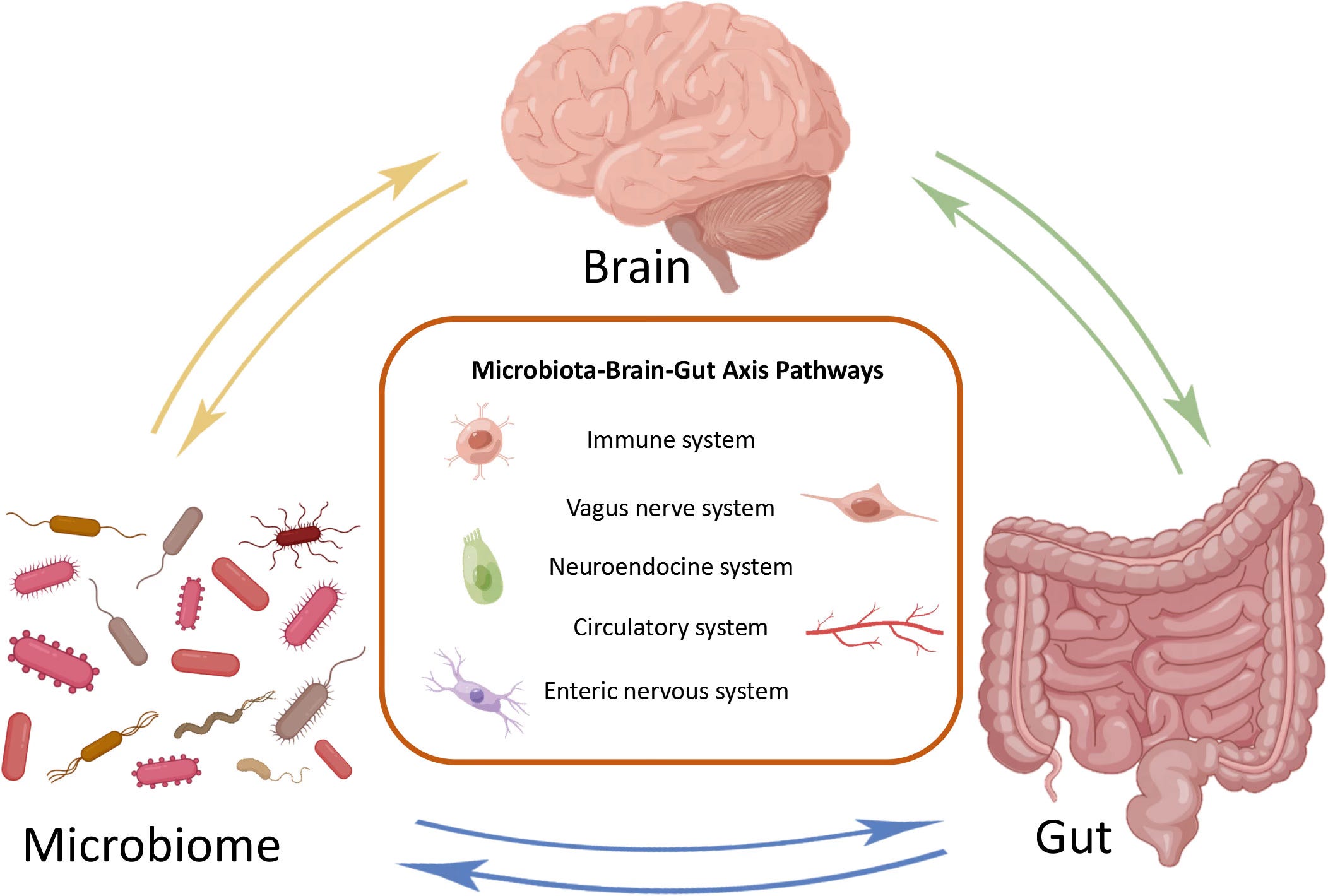
We’ve talked about the vagus nerve here before, so I know you’er already familiar wieth its bsic architecture and the role its afferent fibers play in carrying sensory information from the body to the brain. Just as a quick refresher before we dig in: about 8-% of its fibers are afferent, meaning the majority of the traffic is traveling upward from the gut rather than downward from the brain (Bonaz et al., 2018), and the gut is one of the richest sources of that sensory input.
Now, vagal afferent fibers are woven throughout the layers of the digestive wall, but they don’t actually cross the epithelial barrier, so they can’t make direct contact with the microorganisms living in the gut lumen. They’re picking up signals through intermediaries instead, and there are two worth knowing about. The first is straightforward: bacterial metabolites like short-chain fatty acids diffuse across the mucosal barrier and reach vagal nerve endings that way. The second is something I found really fascinating when I came across it. Neuropod cells, a specialized type of gut sensory epithelial cell that were only first described in 2015, synapse directly with vagal neurons and transmit luminal signals to the brain using glutamate as their neurotransmitter (Mörkl et al., 2023). Receptors for GABA, serotonin, dopamine, and acetylcholine have all been identified on vagal afferent nerves (Mörkl et al., 2023), and the neurotransmitters we talked about in Part 1 are landing on a neural interface that is built to relay those signals directly to the brain.
Source Unknown
What the Vagotomy Studies Tell Us
This is where the research gets really interesting, and also where it runs into an ethical wall I want to be upfront about.
