


Welcome to Part 2 of our HPA-axis series!
When I started planning this series, I thought we might spend some time looking at all the different body systems the HPA axis manages to get its little hands into. And we still might! But as I was working through the research, something else tickled my fancy: the common belief that chronic stress can cause autoimmune disease.
If you’ve spent any amount of time in wellness spaces, you’ve probably heard some version of this. Stress causes inflammation. Chronic stress raises cortisol. Too much cortisol disrupts the immune system. Eventually, autoimmune disease enters the conversation.
But what is the physiology underneath all of that? I wondered.
That’s what we’re investigating in Part 2! Here! Now!
We’re going to look at how the HPA axis and immune system communicate, what can change when stress becomes chronic, and how researchers are trying to understand the relationship between stress physiology, inflammation, and autoimmunity. Along the way, we’re going to ask:
If cortisol helps control inflammation, why can chronic stress be associated with more inflammation?
So what does cortisol look like in people with autoimmune disease?
I tried linking each question in the table of contents to its section below! So if there’s something you want to learn about ASAP, you should be able to click the question above and jump right to it instead of scrolling through the whole article. :) Let me know if it works for you!

How Does the HPA Axis Communicate With the Immune System?
Cortisol gets talked about so often in the context of stress that we sometimes totally forget it has jobs throughout the body that extend beyond stress! For example, cortisol has a role in helping regulate immune activity. What does that mean? Well, immune cells are responsive to glucocorticoids, including cortisol, which gives the HPA axis a direct line of communication with the immune system (Nunez et al., 2025).
A quick terminology stop before we go any further: glucocorticoids are a class of steroid hormones produced by the adrenal cortex, and cortisol is the primary glucocorticoid in humans. Immune cells express glucocorticoid receptors (GRs), so cortisol released into circulation can reach these cells and bind to receptors inside them. Once cortisol binds to a GR, the receptor changes shape, and the cortisol-GR complex can move into the nucleus of the cell, where it can influence gene transcription (Cohen et al., 2012).
This is one of the ways an endocrine signal from the HPA axis can become a change in immune-cell behavior!
One place we can see this is in the regulation of cytokines. Cytokines are signaling proteins that immune cells use to communicate, and some of them help initiate and coordinate inflammation. Glucocorticoid signaling can reduce the production of pro-inflammatory cytokines such as IL-1β, IL-6, and TNF-α (Nunez et al., 2025).
There’s some really interesting molecular biology underneath that effect, involving inflammatory transcription factors such as NF-κB and AP-1. Keep those in your back pocket for right now because we’ll be getting into them a bit later on (Cohen et al., 2012).
Cortisol can also influence where immune cells go throughout the body, which I think is such a cool part of this physiology. This is called leukocyte trafficking. White blood cells are constantly moving between the bloodstream and tissues, and glucocorticoids can change that distribution (Nunez et al., 2025). One example is neutrophil demargination. Some neutrophils are temporarily associated with blood-vessel walls rather than freely circulating in the bloodstream, and glucocorticoids can shift more of those cells into the circulating pool (Cohen et al., 2012).
The effects of glucocorticoid signaling extend across several immune-cell populations, including lymphocytes, macrophages, neutrophils, dendritic cells, and natural killer cells. Depending on the cell type, glucocorticoids can influence aspects of immune-cell trafficking, maturation, and function (Nunez et al., 2025). So when we say that cortisol helps regulate immune activity, we’re describing quite a few different physiological processes happening under that umbrella: changes in inflammatory signaling, changes in what immune cells are doing, and changes in where some of those cells go throughout the body.
ALRIGHT SO: the HPA axis communicates with the immune system through glucocorticoid signaling, with cortisol carrying that signal to immune cells throughout the body. Because those cells can respond directly to cortisol, changes in HPA-axis activity have the potential to change immune activity too!
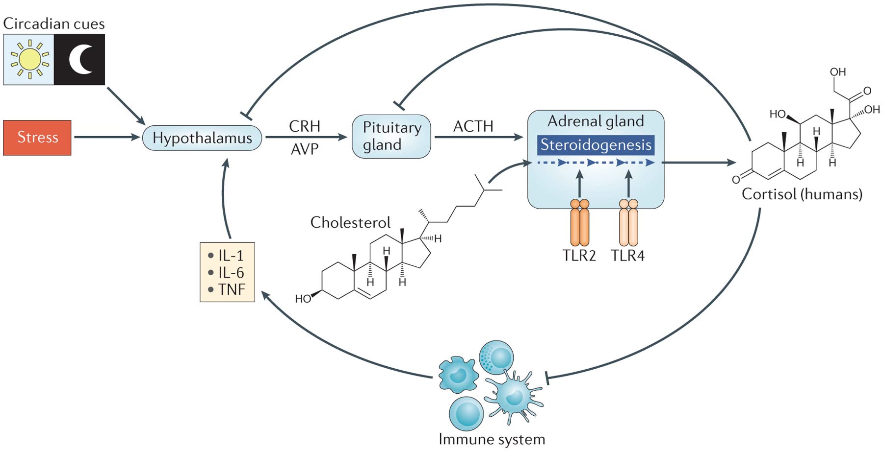
Glucocorticoids help regulate HPA axis activity through negative feedback at the hypothalamus and pituitary while also reducing the expression of pro-inflammatory cytokines.
What Changes When Stress Becomes Chronic?
In Part 1, we spent a lot of time with the negative feedback loop that helps regulate the HPA axis.
A stressor activates the HPA axis, cortisol is released, and rising cortisol eventually feeds back to the hypothalamus and pituitary to help regulate the response. Once the stressor has passed, that feedback helps bring HPA-axis activity back down.
refresh your HPA axis memory here:

But what happens when stress keeps showing up?
With chronic stress, the HPA axis can be recruited repeatedly over time, and eventually the regulation of the system itself can start to change. Researchers have described alterations in the normal pulsatile and circadian patterns of cortisol secretion, changes in negative feedback, and changes in glucocorticoid-receptor sensitivity (Nunez et al., 2025).
I want to hang out with that for just a quick minute because there are several different parts of HPA-axis regulation that we can look at when we talk about “cortisol.” When we’re trying to understand what chronic stress might be doing to this system, we can look at:
How much cortisol is circulating: This is probably the piece we’re most familiar with, but cortisol concentration is only one measurement of HPA-axis activity.
When cortisol is being released: Cortisol follows a circadian rhythm, with concentrations changing throughout the day, and it’s also released in smaller pulses. Chronic stress can disrupt these normal patterns, so two people could theoretically give us the same cortisol measurement at one moment in time while having very different patterns of cortisol secretion across an entire day (Nunez et al., 2025).
How negative feedback is functioning: Cortisol feeds back to the hypothalamus and pituitary, helping regulate further CRH and ACTH secretion. Changes in that feedback can change how the HPA axis responds and regulates itself over time (Nunez et al., 2025).
How sensitive cells are to cortisol: Remember those glucocorticoid receptors from the last section? Okay, perfect. Cortisol has to bind to them for many of its effects on immune cells to occur. Chronic stress has been associated with changes in glucocorticoid-receptor sensitivity, so we also have to consider how responsive those cells are to the cortisol signal they’re receiving. A change in receptor sensitivity could change the biological response to cortisol even when the hormone itself is still circulating (Nunez et al., 2025).
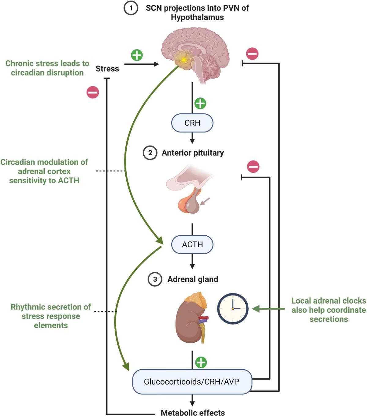
Circadian regulation of the HPA axis and cortisol secretion. The suprachiasmatic nucleus (SCN) helps coordinate daily HPA-axis activity, influencing CRH, ACTH, and glucocorticoid secretion across the circadian cycle. The figure also illustrates how chronic stress can disrupt this rhythmic regulation.
If Cortisol Helps Control Inflammation, Why Can Chronic Stress Be Associated With More Inflammation?
Earlier, we saw that glucocorticoid signaling can reduce the production of pro-inflammatory cytokines such as IL-1β, IL-6, and TNF-α (Nunez et al., 2025). Chronic stress can repeatedly activate the HPA axis, which means cortisol continues to be part of the stress response. So if cortisol is helping regulate inflammation, why can chronic stress be associated with more inflammation?
Back to Those Glucocorticoid Receptors
A big piece of this comes back to those glucocorticoid receptors. During chronic stress, immune cells can become less sensitive to glucocorticoids, a phenomenon called glucocorticoid resistance (Nunez et al., 2025). And to understand why that could change inflammation, you can pull NF-κB and AP-1 back out of your pocket now!
When cortisol binds to its intracellular glucocorticoid receptor, the cortisol-GR complex can move into the nucleus of the cell. There, it can interfere with the activity of nuclear factor kappa B (NF-κB) and activator protein 1 (AP-1), two transcription factors that help regulate the expression of genes involved in producing pro-inflammatory cytokines. Inhibiting their activity is one of the molecular mechanisms through which glucocorticoids can reduce inflammatory signaling (Cohen et al., 2012). The cortisol-GR complex can also interact with glucocorticoid response elements (GREs), which are specific DNA sequences involved in regulating gene transcription. Through this pathway, glucocorticoid signaling can increase the production of anti-inflammatory proteins, including inhibitor of nuclear factor kappa B (IκB) (Cohen et al., 2012).
read more about inflammation & transcription factors here:

IκB gives us a really cool example of what that anti-inflammatory regulation looks like inside the cell as itnbinds NF-κB and keeps it sequestered in the cytoplasm, limiting NF-κB’s ability to enter the nucleus and activate the transcription of inflammatory genes (Cohen et al., 2012). SO! There are multiple points along this pathway where glucocorticoid signaling can influence the inflammatory response.
What Happens With Glucocorticoid Resistance?
Glucocorticoid resistance changes this picture because cortisol can still be circulating and reaching immune cells while those cells have become less responsive to its signal. Reduced glucocorticoid sensitivity can weaken some of the anti-inflammatory effects we just walked through, including the regulation of inflammatory transcription pathways (Cohen et al., 2012).
Human viral-challenge studies give us a really interesting look at this relationship.
In a study of 276 healthy adults, researchers measured chronic psychological stress and glucocorticoid-receptor resistance before exposing participants to a rhinovirus. Greater chronic stress was associated with greater glucocorticoid resistance, and glucocorticoid resistance was associated with a greater likelihood of developing a cold (Cohen et al., 2012).
In a second study of 79 healthy adults, researchers looked more directly at the inflammatory response following rhinovirus exposure. Glucocorticoid resistance measured before infection predicted greater production of IL-1β, TNF-α, and IL-6 afterward. That gives us a mechanism for how chronic stress and increased inflammation can exist in the same physiological picture: if immune cells become less responsive to glucocorticoids, cortisol’s ability to regulate inflammatory signaling can be reduced (Cohen et al., 2012).
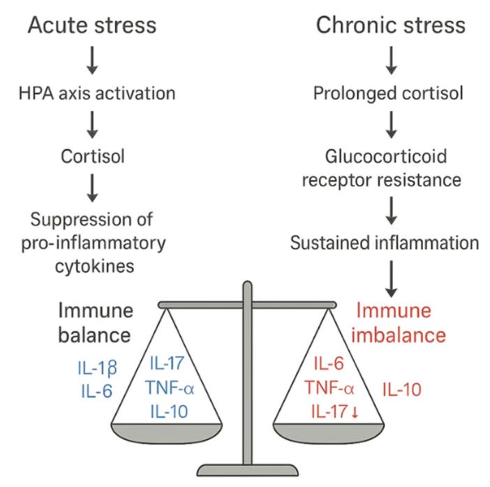
Acute and chronic stress can have different effects on immune regulation. Acute HPA-axis activation and glucocorticoid signaling can help restrain pro-inflammatory cytokine production, while chronic stress may be associated with altered glucocorticoid signaling and glucocorticoid-receptor resistance, reducing this regulatory effect and contributing to sustained inflammation. The “prolonged cortisol” pathway shown here represents one proposed model of chronic-stress dysregulation and should not be interpreted to mean that chronic stress universally produces persistently elevated cortisol (Nunez et al., 2025).
Can the Immune System Change the Stress Response Too?
Alright, so up to this point, we’ve covered how the HPA axis communicates with the immune system, right? Cortisol is released, it reaches immune cells, binds to glucocorticoid receptors, and can change what those cells are doing.
But the immune system gets to pick up the phone and talk back!
“Hello, this is the immune system speaking.”
Part of that return message comes through cytokines. Pro-inflammatory cytokines like IL-1, IL-6, and TNF-α can carry information about inflammatory activity to the brain and activate components of the HPA axis (de Brouwer et al., 2010). That means an active immune response can become part of the signaling that activates the HPA axis and ultimately influences cortisol release. The immune system has officially joined the conversation!
Personally, I think this bidirectional communication gives us a much better way to understand the relationship between stress and inflammation because we have signals moving in both directions! When inflammatory signaling continues over longer periods of time, pro-inflammatory cytokines may also contribute to changes in HPA-axis activity (de Brouwer et al., 2010).
What Does Any of This Have to Do With Autoimmunity?
Okay, now you’re probably like, “Agy, you promised me an article about autoimmunity. CUT TO THE CHASE.”
Immune Tolerance
We’ve talked a LOT about inflammation, but inflammation and autoimmunity aren’t the same thing. Autoimmune diseases involve a loss of immune tolerance toward the body’s own antigens, meaning the immune system begins reacting against components of the body that it would normally recognize as “self” (Nunez et al., 2025).
Immune tolerance is essentially how the immune system learns what belongs to us and what doesn’t. We need an immune system that can recognize and respond to something potentially harmful while maintaining tolerance toward our own tissues. Glucocorticoids can influence this process through their effects on dendritic cells, which are involved in antigen presentation and help shape the immune response that follows. During HPA-axis dysfunction, changes in glucocorticoid signaling may interfere with these effects on dendritic cells and their role in maintaining self-tolerance (Nunez et al., 2025).
learn how this immune tolerance happens in the gut here:

Tregs and Th17 Cells
Regulatory T cells, usually shortened to Tregs, help maintain immune tolerance by suppressing autoreactive immune responses. Under chronic stress and HPA-axis dysfunction, reductions in Treg number and function have been described, along with reductions in IL-10, a cytokine involved in limiting inflammatory responses. T helper 17 cells, or Th17 cells, produce IL-17 and participate in inflammatory immune responses, and increased Th17 activity and IL-17 have also been described in chronic stress and HPA-axis dysfunction (Nunez et al., 2025).
A Bigger Neuroendocrine-Immune Network
There’s another proposed framework I want to bring into this because it widens our view beyond cortisol a bit, which is important.
Researchers have described a neuro-endocrine-immune network in which the nervous, endocrine, and immune systems communicate through hormones, neurotransmitters, neuropeptides, and cytokines. Cortisol is part of that network, along with mediators such as epinephrine, norepinephrine, acetylcholine, substance P, and inflammatory cytokines (Cristal-Lilov et al., 2021).
The basic idea is that these systems normally regulate one another. Stress mediators, autonomic signals, glucocorticoids, and immune mediators participate in feedback relationships. Within the allostatic framework, repeated or prolonged activation can alter those regulatory relationships, and because the system is nonlinear, dysfunction doesn’t necessarily mean that every mediator simply becomes “high” or “low” (Cristal-Lilov et al., 2021).
In the context of autoimmunity, the proposed problem is a loss of coordination between these systems. Neuroendocrine signals may no longer regulate immune activity appropriately for the inflammatory signals being produced, potentially allowing inflammatory and autoreactive immune activity to persist. In rheumatoid arthritis, for example, high levels of inflammatory cytokines would be expected to stimulate the HPA axis, yet cortisol responses have been described as relatively inadequate for the degree of inflammation present (Cristal-Lilov et al., 2021).
This is still a proposed framework. Researchers haven’t established that disruption of this network causes autoimmune disease, or whether some of these changes develop as a consequence of autoimmune inflammation itself.
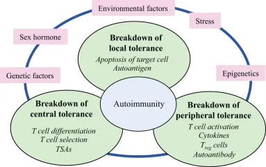
Autoimmunity
So What Does Cortisol Look Like in People With Autoimmune Disease?
The studies show...well, both low and high cortisol?
I know. Hear me out.
In one recent study of people with systemic lupus erythematosus (SLE) and Sjögren’s syndrome, cortisol measured 60 minutes after waking was lower than it was in healthy controls, suggesting a reduced cortisol awakening response and HPA-axis hyporeactivity. Other studies have found higher cortisol exposure in SLE and higher salivary cortisol in Sjögren’s syndrome, while still others have found no significant difference in HPA-axis activity at all (Uçar et al., 2026).
This is why understanding the physiology is SO important.
Cortisol is only one part of the relationship between stress and inflammation. A stressor can activate the HPA axis and lead to cortisol release, but cortisol isn’t required for an inflammatory response to exist. Immune cells have their own receptors, signaling pathways, transcription factors, and cytokines capable of initiating and sustaining inflammatory activity. We’ve already met a few of them in this article: NF-κB, IL-1β, IL-6, and TNF-α. Inflammatory signaling can even travel in the other direction. Pro-inflammatory cytokines including IL-1, IL-6, and TNF-α can communicate inflammatory activity to the brain and activate components of the HPA axis (de Brouwer et al., 2010).
When we measure cortisol in someone with an autoimmune disease, then, we’re looking at one signal within a much larger conversation between the endocrine and immune systems.
There may also be something else driving the inflammatory response.
Autoimmune diseases are multifactorial, with genetic susceptibility, epigenetic changes, environmental exposures, the microbiome, sex hormones, and neuroendocrine stress pathways potentially interacting in their development and activity (Nunez et al., 2025). The neuroendocrine-immune framework, mentioned above, considers how multiple interacting signals, rather than cortisol alone, may shape immune regulation over time (Cristal-Lilov et al., 2021).
So, a lower cortisol measurement doesn’t automatically mean less inflammation, and a higher measurement doesn’t automatically tell us cortisol caused the inflammation.

THAT WAS A LOT. I’m sorry! But I’m not, because now you know :)
We covered a TON of physiology here (if you have to go back and read it, totally fine. My eyes are crossing editing this lolol) but I think it leaves us with a much better way to think about the relationship between chronic stress, cortisol, inflammation, and autoimmunity. The HPA axis and immune system are constantly communicating, and chronic stress can change how that communication is regulated.
Once we widen the lens to the larger neuroendocrine-immune network, it also becomes pretty clear why looking at cortisol alone can only tell us so much.
Cortisol can absolutely be one piece of the puzzle. Its secretion patterns can change, glucocorticoid receptors can become less responsive, and glucocorticoid signaling can influence inflammatory pathways and immune regulation. At the same time, immune signals can influence the HPA axis, and autoimmune disease develops within a much larger landscape of genetic susceptibility, immune tolerance, environmental exposures, hormones, and other interacting factors.
So, eek, don’t hate me but I totally think we can give cortisol a little bit of a break as the end all, be all villain.
A high cortisol measurement doesn’t tell us that cortisol caused someone’s inflammation, and a low measurement doesn’t mean inflammation should disappear. The physiology happening around that measurement is a whole lot bigger than one hormone.
Up next, we’re getting into adaptogens!
if you want to read how I went on citation hunt on this topic, read this:

In the final part of this series, we’ll look at what adaptogens are, where they may interact with the stress-response systems we’ve been learning about, and whether they’re really as safe as we’ve been told they are. :)
Until next time,
— Agy | The Buffalo Herbalist & The Herbal Academic
If you enjoyed this article and want to support the work I’m doing here, you can become a paid subscriber. Paid subscriptions help me continue spending the frankly (lolol) unreasonable amount of time I do reading research papers, following citation trails, and turning all of that into articles like this one.
There’s never any pressure to upgrade, though! Reading, sharing, commenting, or sending an article to someone who might enjoy it all help support my work too, and I’m very grateful that you’re here. 🌻